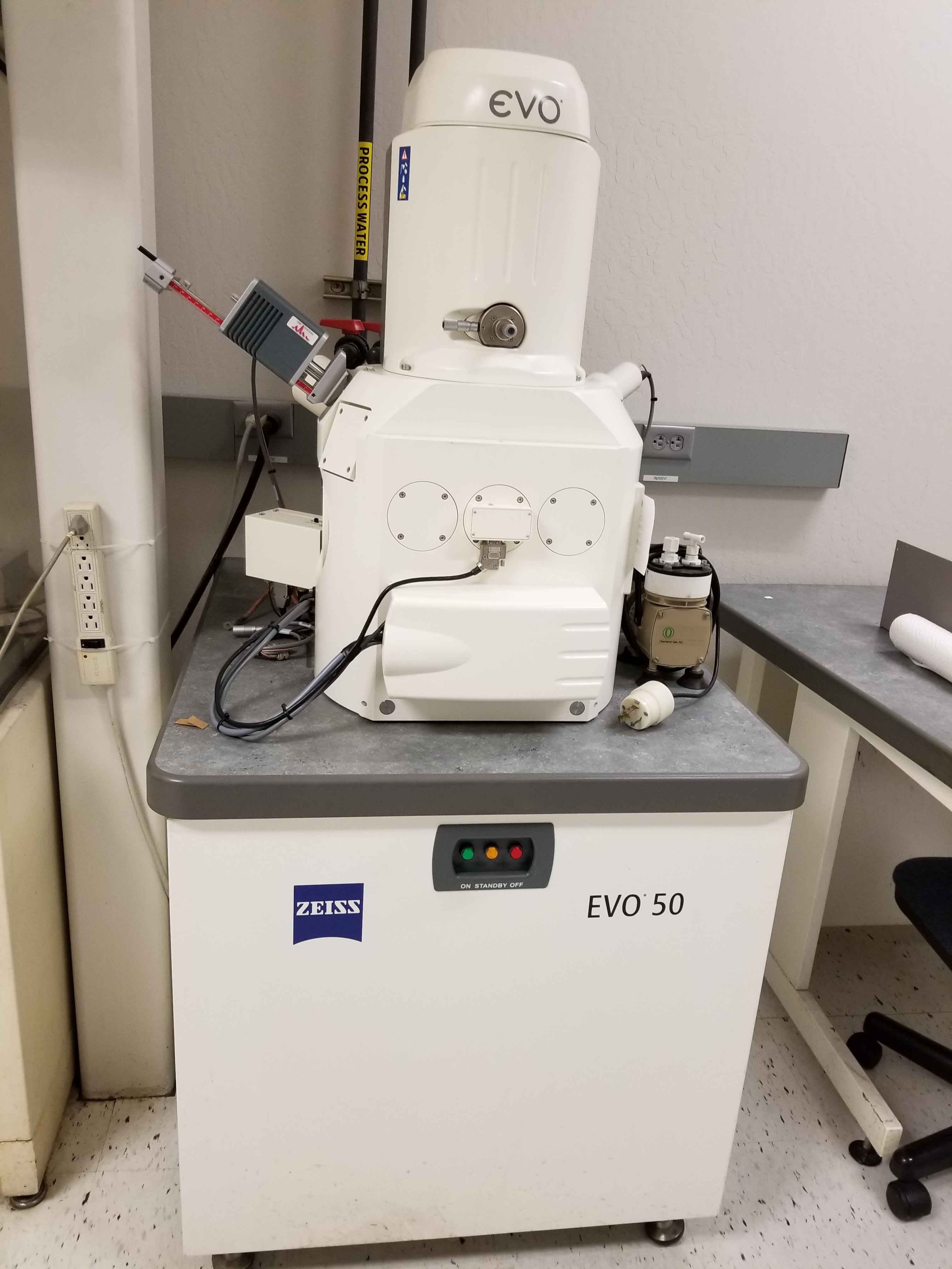
Zeiss Evo 50 Scanning Electron Microscope
AG - EVO 50 Series) scanning electron microscope. Find the very best prices on new and used Zeiss SEM on LabX today. Buy Zeiss products ZEISS EVO MA Series Scanning Electron Microscope New Products. A Miller Dynasty 350 GTA welding power source was used for the manual surfaces of the tested specimens were examined using a Zeiss. The EVO HD cryoSEM, built by Zeiss and Quorum technologies, allows high magnification imaging of cryo-preserved tissue in full vacuum conditions. Magnifications routinely achieved are between 90x and 50,000x with maximum magnification of approximately 300,000x. Zeiss EVO HD15 Scanning Electron Microscope Sainsbury Laboratory Facilities and services. The EVO HD cryoSEM, built by Zeiss and Quorum technologies, allows high magnification imaging of. Magnifications routinely achieved are between 90x and 50,000x with maximum magnification of approximately 300,000x. Excellent contrast is maintained.
ZEISS in Cambridge celebrates 50 years of scanning electron microscopyZEISS proudly celebrates the 50th anniversary of commercial scanning electron microscopy (SEM). In 1965 the first commercial SEM was built by the Cambridge Instrument Company, a UK based predecessor company of ZEISS Microscopy.

Five decades after the launch of the Stereoscan, the ‘industrial gene pool’ of this globally successful technology is evident in a broad range of industry leading SEMs from ZEISS, many of which are still manufactured in Cambridge.The first commercial SEM being demonstrated to Prince Philip, with Sir Charles Oatley. Courtesy of the Royal Microscopical Society, UK.The rise of the scanning electron microscopeAn indispensable toolWith 50 years of continuous growth and development, the SEM has become ever more versatile, and is now firmly established as an indispensable tool for advanced research across many scientific disciplines.
Zeiss Evo 50 Scanning Electron Microscope Parts
With new industrial applications emerging every year, the SEM is also a vital part of research & development, quality control and analysis across an extensive range of industries around the globe.From its early home in materials science, the SEM established itself in the disciplines of electronics, forensics and archaeology. Over the years it has evolved into a key tool for pharmaceutical researchers, food technologists, biologists, geologists and petrologists in the natural resources industries, adapting subtly to fulfil the discrete requirements of each individual application. Use of SEMs for process control and failure analysis is now widespread, with routine SEM analysis an integral part of the semiconductor and automotive industries, and manufacturing more broadly.Coloured electron microscopy of the base of the antenna of a bee. ZEISS EVO SEMAdvances in resolutionSince the introduction of the first commercial SEM, ongoing development has ensured regular advances in resolution. In 1965 The Stereoscan Mark 1 was capable of 10 nanometre resolution. The latest SEM manufactured at the ZEISS factory in Cambridge is the recently launched. Some of the engineers still working at the Cambridge site remember working on the Stereoscans of the late 1960s.
Final Test Engineer Alan Holder, who is due to retire next year, began his career with the Cambridge Instrument Company as an apprentice in 1967. Alan fondly remembers the excitement of the early days, saying “everyone felt that the SEMs we were building had the potential to make a great impact to the world of science.”Coloured electron microscopy of a heavily tilted circuit board from a BSD pre-amp.
The EVO HD cryoSEM, built by Zeiss and Quorum technologies, allows high magnification imaging ofcryo-preserved tissue in full vacuum conditions. Magnifications routinely achieved are between 90x and 50,000x with maximum magnification of approximately 300,000x. Excellent contrast is maintained through the use of a built-in platinum sputter coater enabling user-defined coating on the nanometre scale.
Zeiss Evo 50 Scanning Electron Microscope Diagram


Fracture tools enable fractures through organs and cells revealing delicate structures in the cell walls and membranes. Sublimation protocols enable removal of surface ice or reduction of cytoplasm to reveal organelles.
The time between plunge-freezing and imaging takes as little as 15 minutes.Specifications:Filament: LaB6Detectors: SE, VPSE G3, EPSE, BSDStage: (standard SEM mode) 9 place stage or single specimen Debien Coolstage. For cryoSEM a cryo stage maintains samples at –145C with an anti contaminator maintained at –175C.Cryo prep-deck: maintained at –145C with two fracture knives, platinum sputter coater with thickness monitor. Associated workstation for plunge freezing of fresh tissue with cryo-transfer under vacuum.Access and technical support:cryoSEM drop-in sessions, where users bring samples for cryo preparation and imaging by the facility manager, run from Wednesday 1pm to Friday evening. Please contact the facility manager to reserve a place.
This Site Uses CookiesWe may use cookies to record some preference settings and to analyse howyou use our web site. We may also use external analysis systems which mayset additional cookies to perform their analysis.These cookies (and anyothers in use) are detailed in our site privacy and cookie policies and areintegral to our web site. You can delete or disable these cookies in yourweb browser if you wish but then our site may not work correctly. I have read and understood this message. Hide this message.